X-Ray
Principle
X-rays utilize the penetrating ability of high-energy electromagnetic waves to image human tissues.
Different tissues absorb X-rays at varying degrees. Dense bone tissue absorbs the most, appearing white on film or digital detectors; soft tissues and fat absorb less, appearing gray; gas-filled structures like the lungs absorb the least, resulting in black areas on the final image.
Applications
X-ray examinations are commonly used to assess the following bone structures and related conditions:
Bone Fractures and Dislocations: Clearly show fracture lines, the location of bone fragments, and joint misalignment caused by dislocation. Essential in emergency and orthopedic care.
Bone Degeneration: Detect and assess the degree of bone mineral loss, aiding in the diagnosis and monitoring of osteoporosis. Also observes degenerative changes like bone spurs.
Joint Space Narrowing: Reveal the extent of joint cartilage wear leading to joint space narrowing, assisting in identifying types and stages of arthritis.
Misalignment: Clearly visualize structural abnormalities such as scoliosis, joint displacement, or implant malposition.
Tumors: Initial screening for primary or metastatic tumors such as osteosarcoma, showing lytic or sclerotic lesions’ location and contour.
Infections: Identify pathological signs of infections like osteomyelitis within bones.
")
CT Scan (Computed Tomography)
Principle
The core principle involves capturing multi-angle X-ray images around a specific body part. Unlike standard X-rays, the CT’s X-ray source and detectors rotate around the patient.
Detectors capture attenuated X-ray signals from various angles. These vast amounts of data are then processed by specialized computer systems using complex algorithms to reconstruct high-resolution, multi-angle 2D cross-sectional images or 3D renderings, significantly enhancing contrast and detail resolution.
Applications
CT imaging is suitable for evaluating various complex pathological conditions and internal structures, especially effective for visualizing soft tissues and blood vessels:
Thrombosis: Accurately locates blood clots in the vascular system and assesses their extent.
Subtle Fractures Invisible on X-rays: CT scans clearly show minor or hidden fractures within bones and joints often undetectable by standard X-rays.
Cancer: Precisely delineates tumor size, shape, internal structure, and spatial relationship with adjacent tissues and vessels.
Traumatic Organ and Soft Tissue Injuries: Rapidly diagnoses and quantifies damage such as organ lacerations, contusions, hematomas, and bleeding, also providing crucial insights into deep soft tissue injuries.
Appendicitis: Clearly shows appendiceal thickening, swelling, wall changes, and surrounding fat inflammation.
Infections: Identifies lung inflammation from infections, pulmonary abscesses, deep tissue cellulitis, and complex abscess morphology.
Musculoskeletal Diseases: Reveals complex bone lesions, intra-articular changes, spinal degeneration, and bone sclerosis.
Cardiovascular Diseases: Evaluates coronary artery stenosis, calcification, valve structure and function, large vessel anomalies, and cardiac and pericardial conditions.
Thoracic and Pulmonary Conditions: Detects lung nodules, cancer staging, pneumonia extent, interstitial lung disease, pleural effusion, pneumothorax, and mediastinal lymphadenopathy.

Magnetic Resonance Imaging (MRI)
Principle
MRI uses strong magnetic fields and radiofrequency pulses to detect hydrogen atoms (mainly in water molecules) within the body.
When placed in a magnetic field, hydrogen atoms align in the same direction. Radiofrequency pulses then momentarily disrupt this alignment. As atoms realign, they release weak energy signals, which are captured and processed into highly detailed images.
Because tissues (muscle, fat, water, bone) contain different amounts of water/fat and structural properties, their signal intensity and timing vary, producing highly contrasting images of soft tissues.
Applications
MRI excels in soft tissue visualization and is ideal for diagnosing:
Soft Tissue Injuries of Ligaments, Tendons, Muscles, or Cartilage:
Provides detailed assessment of ligament tears, tendonopathies, muscle strains/tears/hematomas, and cartilage delamination, deep defects, softening, meniscal tears, or labral injuries.
Joint Inflammation: Sensitive to early arthritis changes including synovial inflammation, joint effusion, surrounding bone marrow edema, and later-stage cartilage and bone erosion—essential in assessing disease activity in rheumatoid arthritis and ankylosing spondylitis.
Cartilage Loss: Accurately maps cartilage thickness changes, micro-defects (layering, fissuring), subchondral bone integrity, and edema associated with cartilage wear.
Nerve Compression: Visualizes disc herniations (bulging, protrusion, extrusion, sequestration) and their impact on nerve roots. Clearly displays foraminal narrowing and adjacent anatomical changes.
Spinal Cord Injuries: Detects acute spinal cord contusions, transections, edema; chronic atrophy, softening, or syrinx formation. Also assesses compressive lesions (tumors, hematomas, bone fragments) directly impacting the spinal cord.

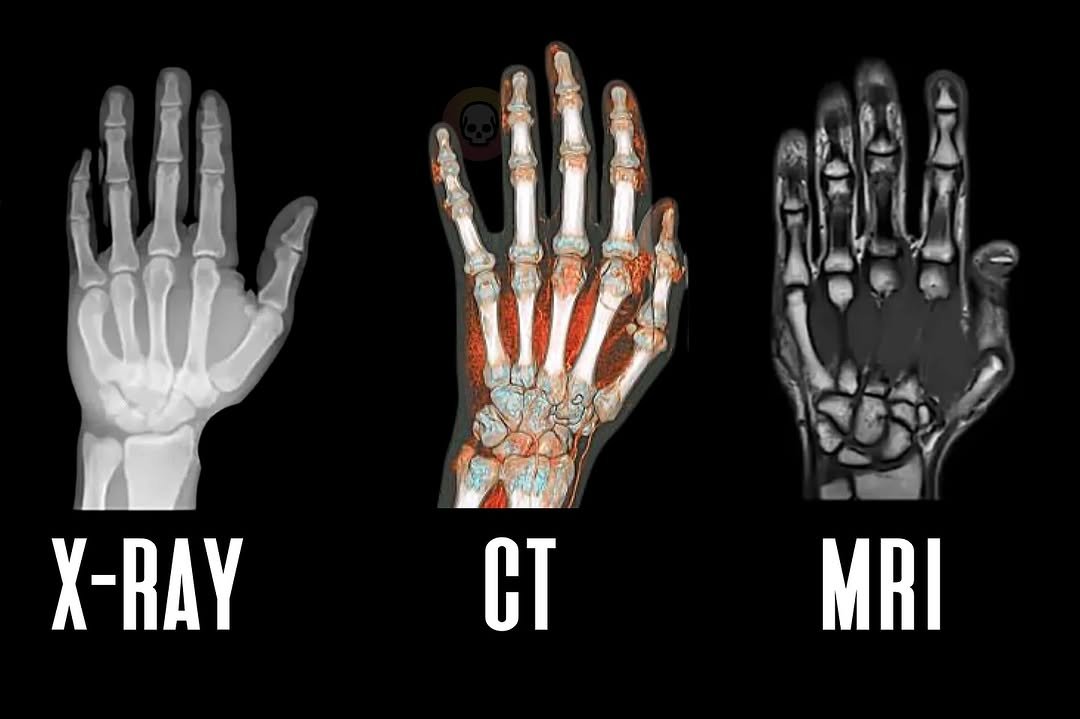
X-Ray, CT Scan, and MRI: Similarities and Differences
Similarities
Despite notable differences, all three imaging techniques share a common goal: to visualize internal structures through the body surface for medical diagnosis. They all produce digital medical images for professional interpretation.
Key Differences
X-Ray
X-rays project a narrow beam through the body, with different tissues absorbing radiation based on their density, creating a 2D image. It uses radiation to generate images. Common applications include detecting fractures, cancer, and pneumonia involving dense tissues like bones or lungs.
Advantages:
- Quick and Convenient:Typically completed within minutes; equipment is easy to operate.
- Low Cost: Generally the most affordable among the three.
- Excellent for Bones:Produces clear images of bones, fractures, and high-density foreign objects.
Disadvantages:
- Ionizing Radiation:Involves exposure to radiation, with cumulative risk.
- Limited Soft Tissue Detail:Poor differentiation of muscles, organs, or ligaments.
- 2D Imaging: Produces overlapping single-plane images lacking depth information.
CT Scan
Essentially a more advanced form of X-ray. The rotating X-ray tube collects projection data from multiple angles. The computer reconstructs fine 3D images and full 360-degree views. CT also uses radiation. It is superior for viewing organs and soft tissues, especially in emergency trauma and vascular imaging.
Advantages:
- Detailed Cross-Sectional Images: Offers axial views of internal structures, minimizing overlapping interference.
- Rapid Scanning: Modern CT systems complete most scans in seconds, ideal for emergencies.
- Versatile:Applicable to almost all body areas and a wide range of conditions.
Disadvantages:
- Ionizing Radiation:Higher doses than X-rays, requiring careful risk-benefit assessment.
- Cost: Much higher than standard X-rays.
- Soft Tissue Contrast: Better than X-rays but inferior to MRI for similar-density tissues.
MRI
MRI uses a fundamentally different principle. It relies on magnetic fields and radio waves—not radiation—to induce signal release from tissues, creating detailed 3D and cross-sectional images. It targets hydrogen in water molecules and excels in visualizing the brain, spinal cord, neck, breasts, abdomen, and muscles.
Advantages:
- No Ionizing Radiation: Involves only magnetic fields and radio waves—no radiation risk.
- High-Contrast Images: Exceptional at differentiating soft tissues (e.g., healthy vs. diseased brain tissue, muscle vs. tendon damage).
- Multiplanar Imaging: Easily produces sagittal, coronal, axial, and other sectional images.
Disadvantages:
- Expensive: Highest cost in terms of equipment, maintenance, and procedures.
- Longer Exam Time:Single scans may take tens of minutes to over an hour.
- Not Suitable for All Patients: Magnetic fields contraindicate patients with metal implants (e.g., pacemakers, aneurysm clips), embedded shrapnel, or severe claustrophobia. Noise levels are also significant.